Anatomy and development of the Oral Cavity
The mouth is a hollow cavity that allows food and air to enter the body and has several different functions.
-
Start the processing of food through the many organs in it, that work together
-
Secondary respiratory channel
-
Sound modification and speech production
The mouth is divided into two sections – the vestibule, the area between the cheeks and teeth, and the oral cavity proper, which is mostly occupied by the tongue. The main structures of the mouth are the teeth (tearing and grinding ingested food to make it digestible), the tongue (positions and mixes food, and has receptors for taste), and the palate (separates the mouth from the nasal cavity, creating separate passages for air and food). All of these, along with the lips, help form speech sounds by modifying the way air passes through the mouth.
Anatomy breakdown

The oral cavity and vestibule are fully lined by mucous membranes containing many small glands that, along with the three pairs of salivary glands, cover the mouth in fluid. This ensures it is kept moist and clear of food or other debris.
Lips – soft, fleshy structures that form the outside border of the mouth opening. The lips are flexible and elastic structures, that contain many collagen and elastin fibers. The outside of the lips is continuous with the skin, while the inside is continuous with the mucous membrane of the mouth.
Cheeks – between the outside and inside epithelium layers of the cheeks are layers of connective tissue, nerves and muscles. The cheek muscles include the buccinator, orbicularis oris and zygomaticus major, which move the lips and cheeks.
Tongue – can move to occupy almost any region in the mouth. The tongue is commonly referred to as a muscle, however it is actually an organ made of epithelium, several skeletal muscles, nerves and connective tissue. It contains many small ridges, called papillae, that help it to grip and move food around in the mouth. Taste buds are placed in the valleys around some of the papillae and produce the sense of taste by detecting chemicals in the food. The tongue can also alter or stop the air flow through the mouth, to produce the sound of various consonants
Hard palate – the roof of the mouth, located at the front. It is formed of the upper jaw’s lower part and the palatine bones and has a domed horseshoe shape.
Gums – soft tissue surrounding the base of the teeth that also protect the bone
How the mouth develops
Mouth formation takes place in the early stages (6-8 weeks) of fetal development. A baby’s mouth is very sensitive and for the first several months, mouthing is how infants learn about their surroundings. Bringing things to their mouths allows them to both smell and taste the item and is how they learn about new things. By around six months of age, a baby’s mouth muscles have become more developed.
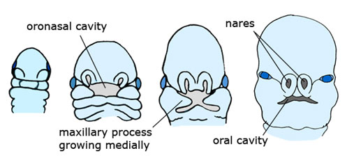
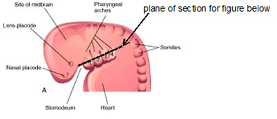
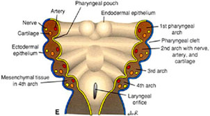
In general, the mouth starts to develop within the first month of pregnancy. Around day 22 (start of fourth week), a set of five structures called the pharyngeal arches are formed. The first arch will develop into the jaws, the second into parts of the face and ears, the third into parts of the hyoid and upper pharynx, and the last two into parts of the larynx and lower pharynx. The formation of the face, the palate and the upper part of the oral cavity depend on the development of the first arch. It splits early into two portions: mandibular (lower) and maxillary (upper) part. By the end of the fourth week, these portions become five distinct swellings: two maxillary, two mandibular and a front midline one, which will form the forehead and the nose. These will continue to swell and eventually fuse to form the external face. A small gap in this fusion becomes the mouth.
Further events of mouth development in pregnancy:
-
At week 7 the baby’s face starts growing.
-
At week 8 the upper lip and nose have formed
-
At week 11 buds for the future teeth appear
-
In the third month the baby can open and close its mouth
-
At week 14 the roof of the baby’s mouth is fully formed, and its constant sucking reflexes are helping to develop its cheeks
A newborn’s tongue is deeply cupped during reflexive sucking.
Fully developed newborns are born with fat pads in their cheeks, usually called sucking pads. These develop during the end of pregnancy. They add stability to the mouth until the baby’s cheek muscles develop.
Tooth anatomy
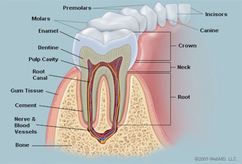
Teeth are calcified structures that serve to break down food. Each tooth is made up of a crown, the exposed portion above the gumline, and a root, which is fixed in the jawbone.
Crown
The crown has a coating of enamel, to protect the dentine beneath.
-
Anatomical crown – the top portion of the tooth and the only part usually visible
-
Enamel – the hardest material in the human body (including bones). It gains this hardness from tightly packed rows of calcium and phosphorous crystals.
-
Dentin – Underneath the enamel is the dentin layer. Dentin is a hard tissue that contains microscopic tubes. When the enamel is damaged, hot or cold air can enter the tooth through these tubes and cause pain.
Neck
-
Gums – also called gingiva, the pink tissue around the neck of the tooth
-
Pulp – innermost portion of the tooth, a soft structure made up of nerves, making it very sensitive, and blood vessels that supply nutrients to the tooth. It also contains lymph vessels that carry white blood cells to the tooth to fight bacteria
-
Pulp cavity – space inside the crown that contains the pulp
Root
-
Root canal – a passageway in the tooth, containing pulp
-
Cementum – Layer of connective tissue that binds the root to the gums and jawbone
-
Periodontal ligament – made of connective tissue and collagen fiber. Helps hold the teeth against the tooth socket in the jaw
The types of teeth are:
-
Incisors – have a somewhat flat crown, a thin edge and a single root. Their main purpose is to cut food
-
Canines – pointed shape and single long roots. Main purpose is to pierce and tear food
-
Molars and premolars – usually have pointed structures on the surface, called cusps. Premolars have 1 or 2 cusps and 1 or 2 roots. Molars have 4-5 cusps and 2-3 roots. Main purpose is to grind and crush food.
How the teeth develop
There are 4 main stages of tooth development
1. At about 6 weeks of pregnancy, the basic substance of the tooth forms in the unborn baby
2. At around 3 to 4 months of pregnancy, the hard tissue that surrounds the teeth is formed
3. After birth, the teeth erupt from the gums
4. The child loses their primary/baby teeth
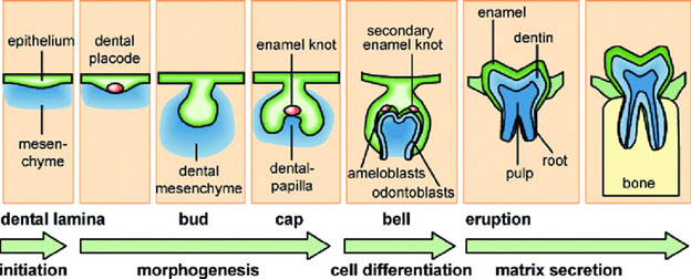
In week 8 of pregnancy, dental buds start to form in the upper and lower jawbone, as the places where milk teeth will grow. In week 10, dental papilla form which will give rise to the dentine and pulp of the milk teeth, with the surrounding epithelium cells beginning to form enamel knots. Permanent tooth buds also begin to develop and are where the permanent teeth will form.
Teething
The breaking of the tooth through the gum line is called eruption. In babies it is called teething. The timing of this differs between children e.g. some children may get teething at only a few months of age, while for others it may not start until they’re 12 months or older.
The usual order of tooth eruption is:
-
Central incisors in lower jaw – between the ages of 6 and 10 months
-
Central incisors in the upper jaw – between 8 and 13 months
-
Lateral incisors in both the upper and lower jaws – between 8 and 16 months, with the lower set usually showing before the upper
-
First set of upper and lower molars – between 13 and 19 months
-
Upper and lower canines – between16 and 23 months
-
Second set of upper and lower molars – between 25 and 33 months
Generally, by the time a child is 3 years old they should have their full set of 20 primary teeth.
Losing primary teeth
From about the age of six onward, baby teeth will start becoming wobbly and fall out, so that the adult teeth can grow. It is also normal for a child to lose their first tooth up to a year or two earlier or later than six years old, with girls usually losing teeth earlier than boys. The first tooth falls out is usually at the front of the lower jaw.
Eruption of permanent teeth
Also known as adult teeth or secondary teeth, the permanent teeth start to develop in the jaws at birth and continue after a child is born. By the age of 21, the average person has 32 permanent teeth – 16 in the upper jaw, 16 in the lower jaw. In some cases, the third molars (or wisdom teeth) don’t develop or don’t erupt, so a set of 28 teeth can be considered normal too. The first permanent molars erupt at roughly 6 years of age (2 in each jaw), behind the child’s existing primary teeth. Other permanent teeth, such as the incisors, canines and premolars erupt into the gaps in the gum left by fallen primary teeth.
The timing for when permanent teeth start showingdiffers with each child. The usual order is:
-
First molars – between 6 and 7 years
-
Central incisors – between 6 and 8 years
-
Lateral incisors – between 7 and 8 years
-
Canine teeth – between 9 and 13 years
-
Premolars – between 9 and 13 years
-
Second molars – between 11 and 13 years
-
Third molars (wisdom teeth) – between 17 and 21 years, if at all